Developmental Biology and Neurogenetics
Prof. Dr. Paul Gottlob Layer

What is Developmental Biology?

Developmental Biology analyses highly integrated feedback processes at molecular, cellular and histological levels, which direct the entire development from fertilisation of the egg through maturation of the adult organism. As systems of study, we use appropriate model embryos, e.g. from chicken, zebrafish or mouse.

This knowledge is ground-breaking for all biomedical progress (e.g. stem cell biology, regenerative medicine, cancer research), as well as for a better understanding of evolution. In this group, we investigate molecular and cellular processes of development of the eyes (retina), the brain, or limbs. Thereby, acetylcholine and its protein components (e.g. cholinesterases) play a central role.

What is Neurogenetics?

Neurogenetics as part of developmental biology investigates molecular bases of the embryonic formation of nervous systems. A general question is, how a huge number of specific neuronal connections as required for a functional brain (1016/person) can be established, given that the total number of genes in man is very limited (2x10⁴/person). Thereby, we analyse roles of acetylcholine and various growth factors.

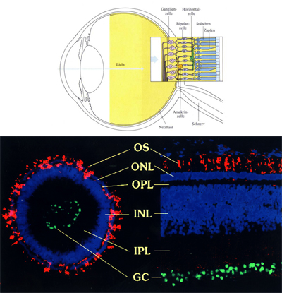
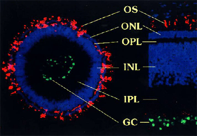
Due to its simple layered structure, we focus on the development of the vertebrate retina in eyes from chicken and mouse embryos. Our cell biological techniques also allow us to culture artificial retinal tissues from stem cells (e.g. retinal spheroids), as well as develop methods for replacement of animal experiments.

Acetylcholinesterase (AChE) degrading the neurotransmitter acetylcholine (ACh) is a most remarkable protein, not only because it is one of the fastest enzymes in nature, but also since it appears in many molecular forms and is regulated by elaborate genetic networks. Moreover, specific inhibitors of cholinesterase (ChEs) play major roles in everyday life as medical therapeutics, as pesticides in agriculture and households, and as chemical warfare agents (nerve gases).

As revealed by sensitive histochemical procedures, AChE is expressed specifically in many tissues during development and in many mature organisms, as well as in healthy and diseased states. Therefore it is not surprising that there has been a long-standing search for additional, “non-classical” functions of cholinesterases (ChEs). In principle, AChE could either act non-enzymatically, e.g. exerting cell adhesive roles, or, alternatively, it could work within the frame of classic cholinergic systems, but in non-neural tissues. AChE might be considered a highly co-opting protein, since possibly it combines such various functions within one molecule.

Our research into ChEs functioning has focused upon i) the expression of ChEs in the neural tube and their close relation to cell proliferation and differentiation, ii) that AChE expression reflects a polycentric brain development, iii) the retina as a model for AChE functioning in neural network formation, and iv) non-neural ChEs in limb development and mature bones. Also, possible roles of AChE in neuritic growth and of cholinergic regulations in stem cells.

AChE functions in proper formation of the mouse retina. An AChE knockout mouse (right) presents disturbed sublayer formation of the inner plexiform layer (IPL; a synaptic layer). Staining: calretinin. Details in Bytyqi et al. 2004.

Possible schemes of developmental actions of ACh and AChE in neurite growth, cell contact formation and cell signaling. (a) secreted ACh from growth cone stops its further advancement („deceleration“). (b) approaching an AChE+ target cell, secreted ACh is degraded and growth cone is further attracted („attraction“), (c) „adhesion“ between two cells could be further stabilised by heterotypic interaction of ChED proteins (e.g. neuroligins, AChE) with neurexins. (d) Alternatively, AChE can bind to laminin of the ECM, which in turn can bind to integrin-2, enabling „adhesion“ and „signaling“ into cell interior. Further see Vogel-Höpker et al. 2010

The highly ordered organisation of cell types within distinct cell layers is a typical feature of many areas of vertebrate brains, representing a necessary requisite of functional neuronal networks. We study the formation of cell layers both in vivo and in vitro, by using as model systems retinae from chick embryos and neonatal rodents.

As our major in vitro approach, we take advantage of so called retinal spheroids, e.g. the cellular reconstruction of more or less complete 3-dimensional retinal tissue spheres from fully dispersed cells. This technology enables us to define the requirements and constraints of the formation of retinal tissues by a cell-to-cell reaggregation analysis. Noticeably, spheres are fully amenable for molecular intervention towards specific aspects of retinal differentiation, e.g. by use of siRNA techniques.

Retinal spheroids can be used i) for retinal tissue engineering, provided that appropriate stem cells become available, and ii) for developing living biosensors (e.g. testing of environmentally dangerous chemicals, such as pesticides), and thus can become valuable tools to reduce animal experimentation.

  • 1948 born in Beutelsbach / Stuttgart, Germany.
  • 1966 Abitur at Gymnasium Waiblingen.
  • 1967 – 1969 Military service with Bundeswehr. Officer candidate. Studies of Russian language.
  • 1969 – 1973 Diploma studies of food sciences in Stuttgart-Hohenheim.
  • 1976 PhD at University of Konstanz under Ferdinand Hucho on photoaffinty labeling of nicotinic cholinergic receptor (AChR) and acetylcholinesterase (AChE).
  • 1977 – 1979 Postdoc at Stanford University, School of Medicine, CA/USA with Eric Shooter. Signaltransduction & Degradation of Nerve growth factor (NGF).
  • 1980 – 1991 Research on development of Vertebrate brains and retina at Max-Planck-Institute for Developmental Biology, Tübingen; (Dpt. Alfred Gierer);
  • 1984 Habilitation in “Zoologie” at University of Tübingen.
  • 1985 – 1990 “Heisenberg” grant award for outstanding research at MPI Tübingen.
  • 1986, 1990 Extended research periods at Institute for Cell Biology Shanghai, leading the first research team of a still ongoing German-Chinese collaboration between the Max-Planck Society (MPG) and Academia Sinica, China.
  • 1991 – 2015 Full Professor and Chair for Developmental Biology & Neurogenetics at Technische Universität Darmstadt (Lehrstuhl at TUDa). Teaching and Research of Developmental, Human & Evolutionary Biology.
  • 1995 – 1996 Serving Director of Institute for Zoology, TUDa.
  • 1999 and 2002 Invited Guest Professor for Cognitive & Brain Sciences, University of Tsukuba, Japan.
  • 1993 – 1994 & 2002 – 2005 Serving as Dean of Faculty for Biology at TUDa (Fachbereich Biologie)
  • 1993 – 2015 Mentor & Chair of Studienstiftung des Deutschen Volkes for most qualified students from all disciplines at TUDa. Award of Daidalos medal in 2013.
  • 2014 Invited Guest Professor for Developmental Biology at Bannari Amman Institute of Technology, Sathyamangalam, Erode, Tamil Nadu 638401, India.
  • 2015 Retirement from teaching & research position.
  • 2015 – 2019 Forschungsmentor of TUDa, supporting young investigators with their grant applications and career planning.

Multiple international cooperations: EU, USA, China, India, Japan. Reviewer for international journals of neuroscience and developmental biology, as well as for many research foundations.

Publication of approx. 200 peer reviewed articles in peer-reviewed journals on the following research topics:

development of retina, brain vesicles, somites, trunk, appendages of chicken embryo and mouse embryos, establishment of retinal spheroid technologies, tissue engineering of retina, non-neuronal embryonic functions of cholinesterases and cholinergic systems, stem cell biology, radiation biology; science philosophy.